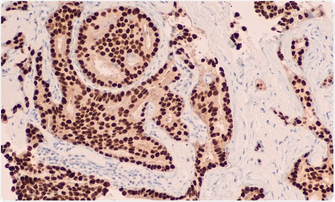
Immunohistochemistry (IHC) is an imaging technique to visualize antigens in cells. Labeled antibodies bind to target antigens in the cell to image the distribution and localization of specific proteins of interest.
David Litman | Shutterstock
The origins of immunohistochemistry (IHC)
The first study using IHC was published in the 1940s when antigens of bacterial species Streptococcus pneumonia were discovered in the tissues. Since then, a wealth of developments and several different approaches have been made. Today, IHC represents a fairly standard routine procedure, but essential in many settings.
Conventional IHC involves staining thin tissue sections immunologically; however, recent techniques can automate the sample preparation and staining. Traditional approaches used one stain or color for one target, but recent developments have furthered the field of multi-color IHC.
Basic methodology
There are generally four steps which precede imaging of the sample: preparation, antigen retrieval, background blocking, and target detection. Because the goal of IHC is to localize cellular components, the tissue architecture and cell morphology needs to be preserved.
In any case, blood must be removed to remove any interference by blood antigens with target antigen detection, while the tissues are fixed, commonly in formalin and then embedded in paraffin wax. The fixation and embedding method is typically referred to FFPE. The embedding allows the tissue to be sectioned and mounted.
Before antigen retrieval, the paraffin from the FFPE needs to be removed to allow the antibodies to react with the antigen. The antigen retrieval itself is done to ensure unmasking antigens which have developed methylene bridges between proteins from the FFPE method. This is typically done by heating the sections or digesting tissue sections.
Some endogenously occurring molecules need to be blocked in IHC to prevent them from giving false positive results. Background blocking involves physically or chemically blocking biotin or enzyme activity. However, background staining can also be caused by nonspecific binding of the antibody. Potential sites are therefore blocked prior to antibody application.
Visualizing cells after IHC
There are two main methods of achieving the visual signal in IHC, called the direct and indirect method. Both direct and indirect methods utilize antibodies to detect the target antigen, but the choice can differ depending on antigen abundance and desired readout.
The direct method has the marker, i.e. the fluorescence or other, labelled on the antibody that binds the antigen. With the indirect method, a secondary antibody is the labeled one, and this secondary one reacts with the primary antibody, which in turn binds the antigen. While the direct method saves time, the indirect method’s signal is amplified, making it much more effective.
The stained samples can be imaged by employing light or fluorescence microscopy. Confocal microscopy can be applied to certain antibody detection methods, and will yield greater detail. In the event of large data sets, high content screening analysis can be used to quickly quantify levels of cellular components and compare data from several samples.
What are the applications of IHC?
IHC is used in the whole spectrum of biological sciences: biological research, diagnosis, and drug development. In biological research, IHC can visualize tissue organization, organ development, and apoptosis, to name just a few fields.
IHC is also the preferred method for studying the expression of genetic material, both mitochondrial and nuclear DNA, in mitochondria in small tissue samples. This is done by antibodies that target DNA or that target the subunits of the respiratory chain complexes encoded by mitochondrial and nuclear DNA.
In medical settings, IHC can be used diagnostically to target specific markers of diseases and conditions. For example, tumor markers can be targeted by IHC to diagnose it as benign or malignant, determine the tumor’s stage, and to identify the cell type.
Finally, in drug development, IHC is used to verify the efficacy of a drug. This can be done by activity detection, or by targeting the up or down regulation of markers of the target disease in the tissue. It can also be used to predict treatment responses when treating different malignancies.
Sources
- www.thermofisher.com/…/overview-immunohistochemistry.html
- https://www.ncbi.nlm.nih.gov/pubmed/17445692
- https://www.ncbi.nlm.nih.gov/pmc/articles/PMC3347652/
- https://www.ncbi.nlm.nih.gov/pmc/articles/PMC3467869/
Last Updated: Jan 3, 2019

Written by
Sara Ryding
Sara is a passionate life sciences writer who specializes in zoology and ornithology. She is currently completing a Ph.D. at Deakin University in Australia which focuses on how the beaks of birds change with global warming.
Source: Read Full Article