Ketamine's long-lasting antidepressant effects unveiled in new study
In a recent study published in the journal Nature, researchers investigate whether persistent blocking of N-methyl-D-aspartate receptor (NMDAR) and burst-type firing by lateral habenula (LHb) neurons may offer a basis for the long-lasting antidepressant effects of an NMDAR agonist, ketamine.
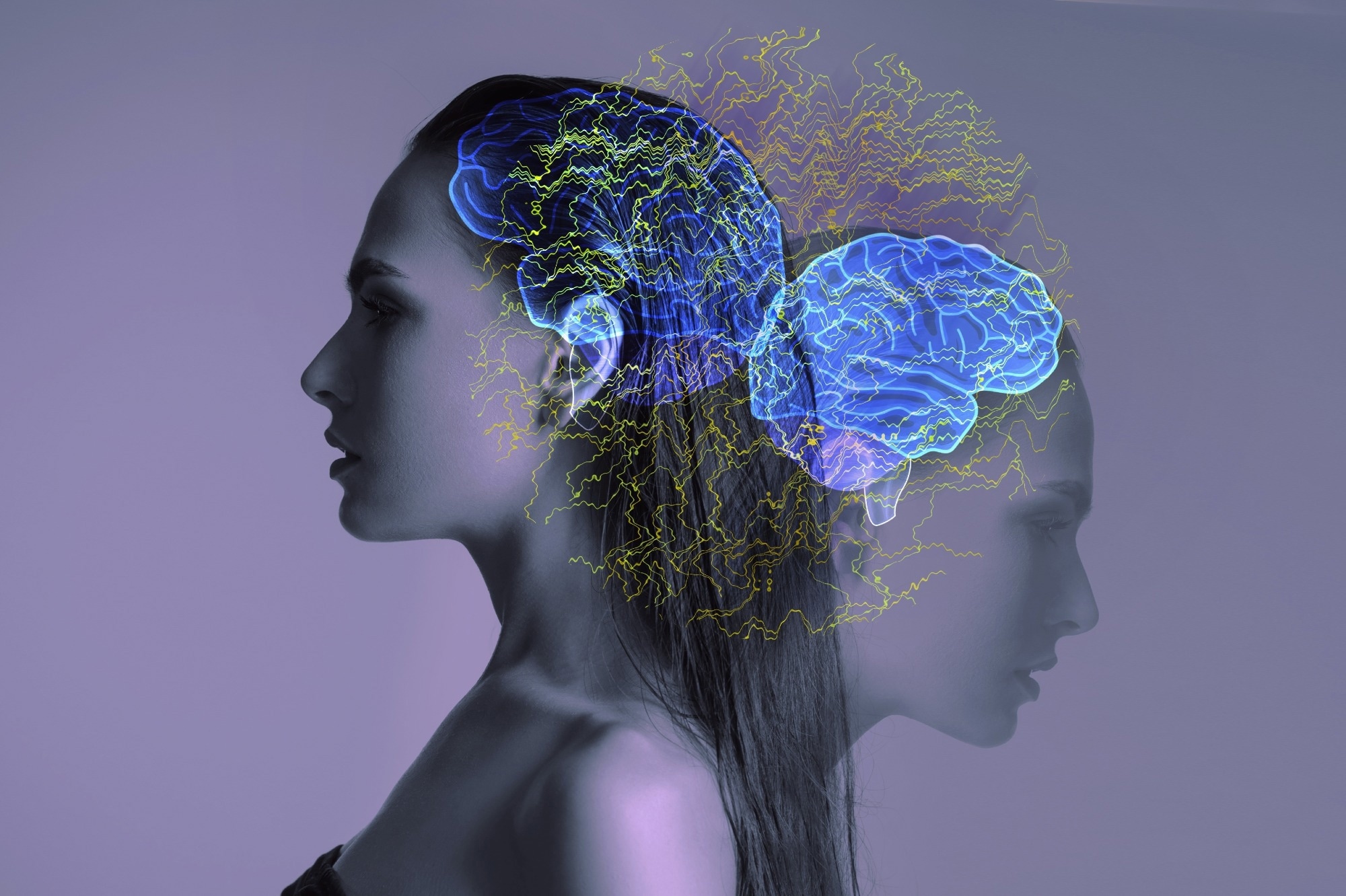
 Study: Sustained antidepressant effect of ketamine through NMDAR trapping in the LHb. Image Credit: Pavlova Yuliia / Shutterstock.com
Background
Ketamine has transformed depression treatment due to its rapid, potent, and prolonged antidepressant effects. Despite having a 13-minute half-life in mice, its antidepressant activity can continue for a minimum of 24 hours.
This has important therapeutic significance, as ketamine inhibits NMDAR-dependent bursting activity in the LHb, which is a hallmark of neuronal activity in several animal models of depression. However, the mechanisms behind ketamine's long-lasting antidepressant effects are poorly known. Furthermore, its long-term effects are not only a fundamental biological concern but also have important therapeutic consequences.
About the study
The antidepressant course of one systemic ketamine injection was examined among chronic restraint stress (CRS) murine models of depression. Mice were administered 10 mg/kg of ketamine intraperitoneally after two weeks of restraint stress. Ketamine concentrations in the brain and plasma were evaluated by liquid chromatography-tandem mass spectrometry (LC-MS/MS) analysis and depressive-like behaviors were assessed.
The researchers investigated whether burst firing in the LHb may explain the long-term effects of ketamine. Spontaneous neuronal activity at rest in coronal LHb brain slices obtained from CRS mice was measured at one hour, 24 hours, or three days following intraperitoneal ketamine treatment. The team subsequently investigated whether a single ketamine injection may cause long-term inhibition of LHb bursting in vivo.
AMPAR- and NMDAR-mediated excitatory postsynaptic currents (AMPAR-eEPSCs and NMDAR-eEPSCs, respectively) were identified in LHb brain slices based on their temporal features and their input-output curves were investigated. Pure NMDAR-eEPSCs were isolated from the LHb of CRS animals using GABAAR blocker, picrotoxin, and the AMPAR blocker, NBQX, to validate the sustained NMDAR blocking effects of ketamine.
The ways in which ketamine would continue to inhibit NMDARs after elimination from the brain were investigated. NMDAR-eEPSC responses in LHb brain slices were constantly measured while washing out ketamine or memantine, a pore-blocking NMDAR inhibitor with a similar affinity but a swifter off-rate than ketamine.
The principal LHb input route, the lateral hypothalamus (LH), was activated to generate LHb burst firing and open NMDARs in vivo. The ability of the 40-Hertz technique to produce real-time place aversion (RTPA) in the animals undergoing the behavioral experiment was examined. The LH-LHb pathway maximized ketamine blockage by opening more NMDARs when ambient levels of ketamine were high.
Study findings
One systemic injection of ketamine suppressed burst-type firing in the lateral habenula by blocking NMDAR receptors for about 24 hours. This long-lasting NMDAR inhibition was related to the usage-based NMDA receptor trapping by ketamine and neural activity controlled the untrapping rate. The researchers took advantage of the dynamic ketamine-NMDA receptor interaction equilibrium by stimulating the LHb and opening local-level NMDA receptors at various ketamine concentrations in plasma.
The FST, but not the SPT, showed an antidepressant tendency three days after the ketamine administration. The antidepressant effects were no longer significant seven days after the injection.
The inherently active LHb neurons were classified as still, tonic, or burst to fire. CRS animals had a considerably higher percentage of neurons that burst-fired than naïve mice. The burst-firing neuronal cell proportion in brain slices prepared one hour after ketamine administration decreased from 40% among saline-treated to 13% among ketamine-treated groups.
The burst-firing neuronal proportion remained much lower 24 hours following ketamine administration at 44% and 24% for saline- and ketamine-treated groups, respectively. The inhibition of LHb bursting activity was non-significant three days following ketamine administration.
Data from both in vitro and in vivo recordings showed that a single systemic ketamine injection in depressive-like animals caused prolonged suppression of LHb bursting activity over a duration that followed its behavioral effects. Likewise, the pharmacologically separated NMDAR-eEPSCs demonstrated considerable inhibition 24 hours after intraperitoneal ketamine administration.
Ketamine binding was influenced by drug concentrations in the environment. In cases of lower ambient concentration of ketamine compared to the dissociation constant (Kd), binding decreased, whereas in situations where it was higher than Kd, binding increased. In vivo, LH-LHb pathway activity at a high ambient ketamine level prolonged its effects on suppressing LHb NMDARs and reduced depression-like behaviors.
Conclusions
The long-lasting antidepressant action of ketamine is likely due to persistent NMDA receptor blockade in the LHb that may be mediated through neural plasticity and other secondary processes. Ketamine could block NMDAR channels for about 24 hours after a single dosage, which is longer than the observed dissociation duration. Furthermore, local ketamine administration in the LHb generated longer-lasting antidepressant effects than systemic administration.
- Ma, S., Chen, M., Jiang, Y., et al. (2023). Sustained antidepressant effect of ketamine through NMDAR trapping in the LHb. Nature. doi:10.1038/s41586-023-06624-1
Posted in: Molecular & Structural Biology | Medical Research News | Medical Condition News | Pharmaceutical News
Tags: Agonist, Antidepressant, Brain, Cell, Chromatography, Chronic, Depression, Hypothalamus, in vitro, in vivo, Ketamine, Liquid Chromatography, Mass Spectrometry, Neurons, Receptor, Spectrometry, Stress

Written by
Pooja Toshniwal Paharia
Dr. based clinical-radiological diagnosis and management of oral lesions and conditions and associated maxillofacial disorders.