Researchers at the University of São Paulo (USP) in Brazil have shown for the first time that stimulating expression of a protein naturally produced by the human body can be a strategy to combat loss of skeletal muscle mass, which happens normally with aging but can intensify in cases of neurodegenerative or inflammatory disease, or in patients who spend long periods in intensive care units (ICUs).
Ten days in an ICU are estimated to result in the loss of up to 20% of muscle mass in the legs and in important respiratory muscles such as the diaphragm. There are no drugs that can effectively treat these cases without severe side-effects. Only physiotherapy, breathing exercises, and electrostimulation can safely reverse ICU-acquired muscle atrophy.
“We were able to demonstrate that overexpression of protein kinase A [PKA] significantly improved muscle resistance to fatigue in mice. This is because PKA both suppresses FoxO proteins, which activate genes associated with atrophy and increases the formation of muscle fibers with augmented oxidative potential [respiratory capacity], thereby promoting hypertrophy and greater resistance to fatigue in specific muscles,” said Luiz Carlos Navegantes, a professor in the Physiology Department of the University of São Paulo’s Ribeirão Preto Medical School (FMRP-USP) and a co-author of the article on the research published in the journal FASEB J.
The study was supported by FAPESP and offers a new direction for the search for drugs that protect muscles from atrophy without severe adverse side-effects such as cardiac hypertrophy, tachycardia, heart attack, and even death.
“The beneficial role of PKA in muscles, which consists of stimulating anabolism and strength, is unique to all known proteins, making it a strategic target and object of study for the treatment of neuromuscular disease and pathological conditions that lead to weakness and muscle atrophy,” Navegantes told Agência FAPESP.
Metabolic relationship
The FMRP-USP research group, which is led by Professor Isis do Carmo Kettelhut, discovered 23 years ago that the hormone adrenalin not only breaks down energy sources such as lipids and carbohydrate but also inhibits degradation of the proteins in muscle fibers. “That was a paradigm shift,” Navegantes said. “Adrenalin had always been considered a hormone that mobilized energy, not one that prevented excessive breaking down of proteins, which would be catastrophic, especially if they maintain muscle contraction.”
Since they discovered that adrenalin protects muscles, the group has investigated the therapeutic potential of drugs similar to the hormone (sympathomimetics) but without managing to avoid adverse side-effects. “Everyone wants to preserve muscle proteins, from athletes who aim to increase muscle mass to patients who need their diaphragm to continue breathing normally,” Navegantes said. “However, these drugs can have effects that damage the organism and may even be deadly because of cardiac hypertrophy.”
The main problem with using synthetic adrenalin and sympathomimetics, he added, is that they have systemic effects and cannot be targeted to specific muscles. This was evidenced in another study conducted by the group in which the sympathomimetic formoterol was tested for chronic treatment of muscle mass loss and fatigue. In an article published in Cachexia, Sarcopenia and Muscle, the researchers described the effects of this drug, which is widely prescribed for asthma, and showed that it can also promote muscle growth and the strengthening of skeletal muscle.
“Muscles grew owing basically to two mechanisms: direct action of the drug inhibiting protein degradation, and increased secretion of insulin, a well-known anabolic hormone,” Navegantes said. “However, although sympathomimetics are extremely interesting to combat atrophy and have already been adopted in clinical practice, they can have adverse side-effects, especially cardiac hypertrophy, and chronic use of these drugs is therefore dangerous.”
Targeted action
The group decided to study PKA as a potential solution to the problem by means of selective gene transfer, targeting a specific muscle. “PKA was known to be activated by adrenalin and hence associated with lipid and carbohydrate breakdown for energy. However, PKA is located inside muscle cells and adrenalin is a circulating hormone. This difference enabled us to target the effect of PKA on a specific muscle in mice.”
In order for adrenalin to inhibit muscle degradation while also breaking down lipids and carbohydrates, PKA must be activated, Navegantes continued. The adrenalin attaches to a cell membrane receptor and sends a signal that leads to stimulation of PKA in the cytosol (intracellular fluid). Suppression of the genes associated with muscle atrophy is one of the final effects mediated by PKA in cell nuclei.
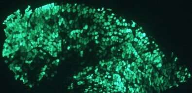
The researchers used electroporation to activate genes associated with PKA production not in all cells but in a single muscle. By this method, a plasmid was inserted in vivo by electric pulse into the muscle, where it modified the DNA of the muscle fiber. Ph.D. candidate Dawit Albieiro Pinheiro Gonçalves learned the method during an internship in Italy with FAPESP’s support.
“It’s a technique used to prove hypotheses in live animals,” Navegantes explained. “You apply a pulse to the muscle you’re studying and introduce a plasmid that modifies the muscle’s genes [by genetic editing] so as to produce a protein of interest, in this case, PKA. In this manner, you can intervene selectively in a specific skeletal muscle without altering the animal’s other tissues.”
The intervention resulted in overexpression of PKA catalytic subunits (PKAcat) and growing oxidative fiber conversion, substantially improving resistance to muscle fatigue. “When we did the opposite, introducing a molecule called PKI that inhibited endogenous PKA, we observed activation of genes associated with atrophy and a reduction in muscle fiber area,” he said.
Source: Read Full Article