Fluorescence spectroscopy is an analytical method based on the fluorescence properties of the sample, and is used for quantitative measurements of drugs, metabolites, and other chemical products.
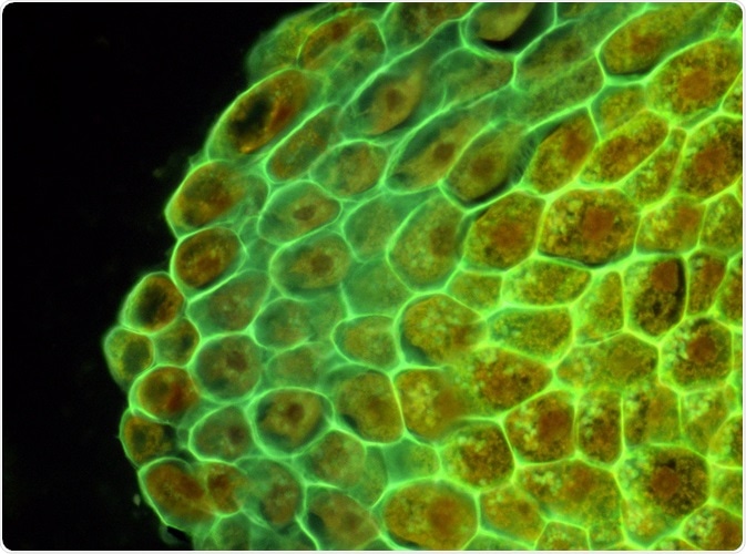 Image Credit: D. Kucharski K. Kucharska
Use of fluorescence
This method uses fluorescence to analyze the samples. In fluorescence, the fluorophore absorbs light of a certain wavelength, which then excites the electron, which moves from ground state to a high energy excited state.
This electron, while coming back to the ground state, emit energy in the form of fluorescence, which is light of a different wavelength. These wavelengths are called the “excitation wavelength” and “emission wavelength”.
As there is energy loss in this change, the light emitted is of longer wavelength that the absorbed light. This difference in the excitation and emission wavelengths is termed as the ‘stokes shift’; if the wavelength of absorbed radiation is ‘a’ and the wavelength of emitted radiation is ‘b’, stokes shift is calculated as ‘b-a’. It is preferred that the stokes shift be higher in fluorophores, as this reduces the interference between excitation and emission spectra.
Intrinsic and extrinsic fluorophores
Intrinsic fluorophores refer to molecules that are naturally fluorescent due to aromatic groups present in the amino acid side-chains. Few examples of such compounds are tyrosine, tryptophan, phenylalanine, and co-factors, such as FMN, FAD, and NAD.
Extrinsic fluorophores are compounds which do not show fluorescence naturally. Fluorescence spectroscopy can be performed on compounds which are not intrinsically fluorescent by attaching a fluorescent probe to these compounds. Some examples of these are fluorescein, ethidium bromide and acridine orange.
Quantum efficiency
Quantum efficiency is an important parameter in fluorescent measurements which provides a measurement of photon vs converted electrons, or the ratio of how many incident photons are absorbed vs the number converted to electrons. This parameter is high is samples at low concentration.
Factors that affect fluorescence spectroscopy
Molecular rigidity
Fluorophores with rigid structures are preferred for performing fluorescence spectroscopy, as they have reduced vibrations and reduced probability of transitioning to triplet state. Fluorescein and eosin have rigid structures and are strongly fluorescent, while phenolphthalein is non-rigid and non-fluorescent.
Solvent polarity
The polarity of the solvent can also dictate the degree of fluorescence. The fluorescence of the structures can decrease in the presence of heavy atoms in the solvent.
Dissolved oxygen
Dissolved oxygen in the solvent can also affect the fluorescence by reducing the emission intensity. This is achieved by photochemical oxidation of fluorophore. The paramagnetic properties of oxygen can also lead to quenching of fluorescence.
pH
pH can affect the fluorescent properties. One such example is aniline, which exists as a cation at lower pH and as anion at higher pH. In both instances, the fluorescence is lost. .
Quenching
Quenching refers to decrease in the fluorescence intensity. This may be caused by absorption of fluorescence by the solution or absorption of the fluorescent material itself. The latter effect is called self-quenching.
Instrumentation
Fluorescence spectroscopy involves the use following components: a light source, monochromator, and a detector. The usual light source includes xenon or mercury arc lamps (300-800 nm).
A monochromator is used to select a specific wavelength from the incident beam which is then incident on the sample. The emission monochromator determines the emission wavelength. Detector refers to a sensitive photomultiplier which detect and collects the emission wavelength.
Applications of fluorescence spectroscopy
- Fluorescent probes – This method is used to qualitatively and quantitatively detect biological compounds present in low concentration.
- Protein structure – There are several intrinsic fluorophores present in proteins, such as tryptophan, tyrosine, phenylalanine among others. The fluorescence of a protein is determined by all its amino acids, solvent properties, and presence of co-factors, such as NAD, FAD. Thus, this method can help in determining the protein structure.
- Fluorescence-activated cell sorting (FACS) – This is method used to sort a mixture of cells based on the differences in fluorescent properties.
Disadvantages of fluorescence spectroscopy
The fluorescence can be susceptible to different conditions mentioned above, such as pH, temperature, or solvent. Other interfering materials, including detergents, filter paper, and tissue material may also alter and affect the fluorescence. Fluorescence may also be lost by interaction with other molecules and especially in concentrated samples.
Sources:
- Fluorescence spectroscopy as an Analytical Tool.
- Fluorescence spectroscopy.
- Fluorescence spectroscopy.
Further Reading
- All Fluorescence Content
- How do Epifluorescence Microscopes Work?
- GFP-tagging in Fluorescence Microscopy
- Fluorescence Quenching
- Importance of Colocalization Studies
Last Updated: Oct 11, 2018

Written by
Dr. Surat P
Dr. Surat graduated with a Ph.D. in Cell Biology and Mechanobiology from the Tata Institute of Fundamental Research (Mumbai, India) in 2016. Prior to her Ph.D., Surat studied for a Bachelor of Science (B.Sc.) degree in Zoology, during which she was the recipient of anIndian Academy of SciencesSummer Fellowship to study the proteins involved in AIDs. She produces feature articles on a wide range of topics, such as medical ethics, data manipulation, pseudoscience and superstition, education, and human evolution. She is passionate about science communication and writes articles covering all areas of the life sciences.
Source: Read Full Article