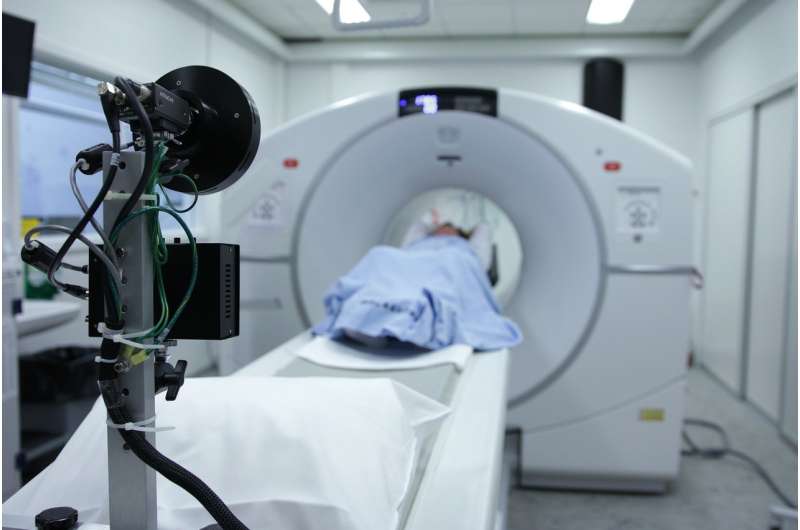
One of the primary tools that oncologists use to stage cancers is the PET (positron emission tomography) scan, an imaging test that uses a small amount of radioactive sugar to detect metabolically active areas within the body.
CU Cancer Center member Sarah Milgrom, MD, recently conducted research to study if PET scans can help to predict recurrence in Hodgkin lymphoma. She used the images to measure the lymphoma volume for each patient at the time of diagnosis, then followed the patients over time to find out whether the initial metabolic tumor volume was associated with the risk of the cancer coming back. Her findings, which led to her being selected for a Young Investigator Award from the Children’s Oncology Group, can help cancer doctors more accurately predict patients’ risk of recurrence, which, in turn, may help them to choose the most appropriate treatments.
We talked with Milgrom about her research and what it means for patients.
Q: How do PET scans work?
A: Prior to the scan, a small amount of a radioactive sugar is injected into a vein. This sugar, or radiotracer, collects in areas of the body that are metabolically active, since they take up a lot of sugar. Then, the PET scanner creates detailed pictures of the body, showing areas that have absorbed large amounts of the radiotracer. PET scans are used commonly in many types of cancers, including Hodgkin lymphoma. Hodgkin lymphoma cells take up more sugar than normal cells, so they are detected by PET scans. In this way, PET scans can pinpoint the locations of the lymphoma within the body.
Q: What was your hypothesis regarding PET scans in your study on metabolic tumor volume in children and adolescents with intermediate-risk Hodgkin lymphoma?
A: We wanted to see if the volume of disease in a patient’s body at the time of their diagnosis was associated with their risk of recurrence after treatment. For this study, we looked at patients with intermediate-risk Hodgkin lymphoma who were treated on a Children’s Oncology Group (COG) study. We only included patients who received the same chemotherapy and radiation treatment regimen. We used PET scans to measure the lymphoma volume for each patient at the time of diagnosis, and we explored whether the initial metabolic tumor volume was associated with the risk of cancer recurrence.
Q: What were your findings?
A: We found that a higher volume of lymphoma at diagnosis was associated with a greater risk of recurrence after treatment. Importantly, metabolic tumor volume was associated with the risk of recurrence even when we controlled for other important variables like stage, bulk, and response to chemotherapy. These are the risk factors that are used currently to select the appropriate treatment for patients. This means that metabolic tumor volume might add prognostic information on top of standard risk factors, so it could be used to help select the best treatment for patients.
Q; What will this research mean for patients going forward?
A: In the future, it is possible that measures of baseline metabolic tumor volume will be used to select the most appropriate treatment for patients with intermediate-risk Hodgkin lymphoma. This could lead to improved patient outcomes.
Q: You did a similar study in children with high-risk Hodgkin lymphoma. How did that one work?
A: That study included patients who were treated on a different COG protocol that was for patients with high-risk Hodgkin lymphoma. On that protocol, patients received different treatments, depending upon how quickly their disease responded to chemotherapy. Patients whose disease had responded very well after the first two cycles of chemotherapy were called “rapid early responders,” and they received less intensive therapy than patients whose disease had not responded as well at that time (“slow early responders”). Again, we used PET scans to measure the initial lymphoma volume for each patient to explore whether that volume was associated with the risk of cancer recurrence. First, we evaluated the entire group, and then we looked at the rapid early responders and the slow early responders separately.
Q: What were your findings in the study on high-risk patients?
A: We found that total lesion glycolysis—a measurement that considers the total body volume of disease as well as its metabolic activity—was significantly associated with the risk of recurrence in the total cohort. When we looked at the rapid early responders and the slow early responders separately, however, we had a very interesting finding. Total lesion glycosis was associated with the risk of recurrence in rapid early responders, but not in slow early responders.
Rapid early responders with a low baseline tumor burden experienced excellent outcomes with less intensive therapy; however, rapid early responders with a high baseline tumor burden experienced poor outcomes that were even worse than those of slow early responders. This suggests that patients with a high upfront tumor burden may benefit from intensified therapy, even if they achieve a rapid early response.
Q: What will your research on high-risk Hodgkin lymphoma mean for patients going forward?
A: PET-based measures of initial disease burden may help us to predict patients’ risk of recurrence. That, in turn, may help us to select the best treatment for patients with high-risk Hodgkin lymphoma.
Q: Will using PET scans to measure initial disease burden change anything about the way doctors care for cancer patients?
Source: Read Full Article