The brain is a sophisticated biological system known to produce different experiences and perceptions via complex dynamics. Different brain regions and neural populations commonly work in tandem, communicating with each other to ultimately produce specific behaviors and sensations.
Researchers at University of Oxford and the Max Planck Institute for Dynamics and Self-Organization recently carried out a study aimed at better understanding the neural dynamics underpinning this communication between neural populations. Their findings, gathered in Nature Neuroscience, show that the probability that mice will perceive something is linked to a variability of neural activity in the brain region that processes the incoming stimulus information.
“Generally, we are interested in how the brain processes information,” James Rowland and Thijs Van der Plas, co-authors of the paper, told Medical Xpress. “The brain receives inputs from the senses which reflect what is happening in the world around it. It must then make sense of this information and use it to make decisions and take actions. To achieve this, the brain is built on a principle of division of labor, where different regions are specialized to perform distinct tasks.”
Previous seminal works have often tried to pin-point how the brain supports the completion of different specialized tasks, such as visual or auditory tasks. These studies gathered some interesting findings, consistently highlighting the existence of distinct neuron populations focused on specific aspects of tasks.
“For example, neurons in the brain region associated with vision activate in response to areas of an image with sharp changes in contrast,” Rowland and Van der Plas explained. “However, at the same time, we know that brain regions do not operate in isolation; to understand the world in full, they must reliably communicate.”
The primary objective of the recent work by Rowland and Van der Plas was to better understand how different brain regions send and receive messages, as well as what these “messages” essentially consist of. To explore these research questions, the researchers carried out a series of experiments on mice, as they are known to have a brain structure that resembles that of humans.
“A crucial way for mice to make sense of the environment is through their whiskers, they use them in the same way we’d feel with our fingertips if we were navigating a dark corridor,” Rowland and Van der Plas said.
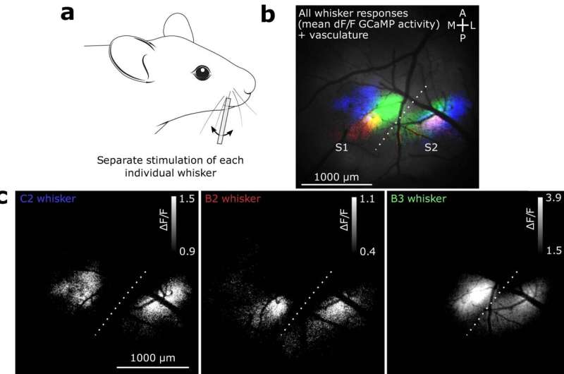
“Their brains reflect this, with a large proportion of their cortex assigned to information coming from the whiskers. To conduct our study, we focused on two specialized regions of the mouse brain (called ‘primary somatosensory cortex’ and ‘secondary somatosensory cortex,’ also known as S1 and S2) that receive inputs from the whiskers.”
When mice are trying to make sense of the world around them, the S1 and S2 regions of their somatosensory cortex have been found to closely communicate with each other in a bi-directional way. In their experiments, the researchers thus decided to focus on these two brain regions.
Rowland, Van der Plas and their colleagues monitored the activity of awake mice while also simultaneously recording neural activity in the S1 and S2 regions. Their recordings were performed using a fluorescence imaging tool known as a two-photon microscope.
“In addition to recording neurons, we used a technique called two-photon photostimulation to artificially manipulate neuronal activity,” Rowland and Van der Plas said.
“This allowed us to send messages directly into S1 and observe under what conditions they were successfully delivered to S2. Finally, we trained mice to perform a behavioral task, which allowed us to ‘ask’ the mice whether or not they had perceived our message sent into S1.”
The experiments performed by the researchers allowed them to ascertain that the messages perceived by the mice were those “sent” by the S1 segment of the somatosensory cortex to S2 or other brain regions. In addition, the team’s findings are aligned with those of previous studies, suggesting that changes in behavior can be prompted by the activity of an extraordinarily low number of neurons.
“Mice have between 50 and 100 million neurons, but we confirmed that inducing activity in just a dozen or so can alter their behavior, an astonishing degree of sensitivity,” Rowland and Van der Plas said.
“We were then able to show that a behavioral response only occurred when this activity was propagated out of the brain region where the signal originated. The second finding is the importance of the ‘signal-to-noise’ ratio of neural activity in communication between brain regions. This is best explained by imagining neurons as people chatting in a bar. It’s much easier for the neurons to communicate when they speak loudly and the bar is quiet than when it’s noisy and they speak quietly.”
Based on their observations, Rowland, Van der Plas and their colleagues hypothesize that the mouse brain, and potentially the human brain too, actively ‘quiets itself’ when it knows that an important message is incoming. This process could be primarily salient in situations where the communication between brain areas can make a huge difference, such as in potentially threatening life or death situations.
This recent work could soon pave the way for further studies in this area. Collectively, these research efforts could help to uncover the underpinnings of communication between different neuronal populations, which could also improve the understanding of mental disorders in which this communication is impaired.
“Further work in this area could focus on the underlying mechanism involved in this ‘quietening down’ process,” Rowland and Van der Plas added. “One hypothesis is that this process could be mediated by a chemical in the brain (known as a neurotransmitter) such as acetylcholine which is known to be disrupted in many neurological diseases, potentially tying our research into clinical research down the line.”
More information:
James M. Rowland et al, Propagation of activity through the cortical hierarchy and perception are determined by neural variability, Nature Neuroscience (2023). DOI: 10.1038/s41593-023-01413-5
Journal information:
Nature Neuroscience
Source: Read Full Article