Mammalian antibodies typically have two arms that can bind to two individual antigen molecules. But what is the optimal antigen distance, and does the doubled antigen-binding region improve antibody binding strength? Scandinavian researchers have recently measured the optimal antigen-distances for different antibody classes by presenting controllable antigen patterns on DNA-origami scaffolds.
Skip to:
- DNA Origami for Construction of Controllable Antigen-patterned Surfaces
- Antibodies Bind Strongest at 16 nm Antigen Distances
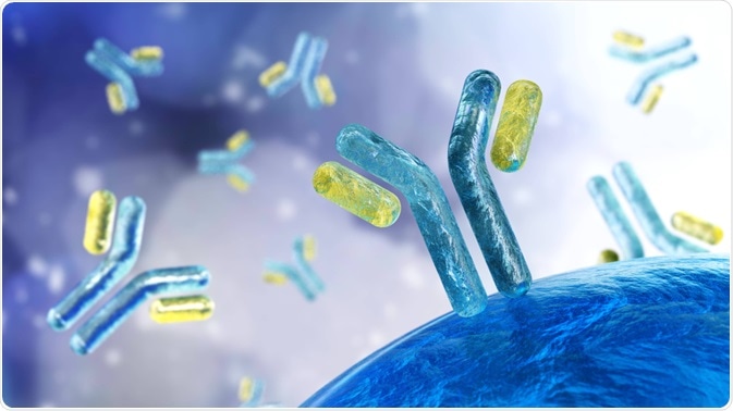 Image Credit: ustas7777777 / Shutterstock.com
The research has been published in January in the Journal Nature Nanotechnology.
Humans have different types of antibodies, such as the Immunoglobulins G (IgG) and M (IgM). IgG is the antibody class which is the most abundant in blood, while IgM is usually the antibody class that first occurs after the immune system is challenged by a new antigen. All these antibodies are characterized by a Y- or T-shape.
These shapes are the result of a dimeric, cross-linked protein (“heavy chain”) which is further associated with another type of protein (“light chain”) on flexible arms of the antibody. The antibody base is rather conserved in amino acid sequence, while the flexible arms are variable and thus make antibodies specific for the binding of different antigens, rendering antibodies effective agents of immune protection.
While each Y- or T-shaped antibody already has a doublet antigen-binding site, the conserved antibody base region can aggregate with other antibody molecules, e.g. into a hexameric antibody assembly with a total of 12 antigen-binding sites.
It is conceivable that when antibody-assemblies with a larger number of binding sites circulate in the blood stream they will be more likely to catch an antigen, or multiple at once. But does this also strengthen the binding of antibodies to surfaces patterned with multiple antigens at various distances?
Previous research has shown that antibodies can kind of “walk” on antigen-patterned surfaces, by alternated binding to and dissociation from antigens, enabling each arm to “step” from one antigen site to another as long as the antigens are at a reasonable distance. The strength of the single- versus double-site binding is not well understood.
Recently, Scandinavian researchers from Stockholm and Oslo, led by Associate Professor Björn Högberg, have demonstrated that IgG and IgM antibodies indeed bind stronger when two antigens are presented on a surface as long as their distance is below 17 nm.
DNA origami
The researchers built patterned surfaces with DNA origami. They used different arrangements of DNA strands, hosting special DNA stretches at known locations where the antigen was attached thereafter. These known locations were at distances of between 3 and 44 nm, and for comparison, a version with only a single binding site was included. The scientists formed the origamis on surfaces suitable for surface plasmon resonance (SPR) spectroscopy, a technique that is commonly used to measure the interaction strength between two biomolecules, such as an antibody and an SPR-surface-immobilized antigen.
The investigators compared the binding strength of different antibody types to their antigens, including antibodies with a known high or low affinity to the same antigen. From the binding data, they constructed a mathematical model to associate the binding strength with different binding modes, i.e. single- versus dual- antigen binding.
When do antibodies bind the strongest?
The scientists could show that all the antibodies tested bound strongest to a surface-bound antigen couple at 16 nm distance. Interestingly, they observed a sharp decrease in binding strength at only a 1 nm longer distance, i.e. 17 nm, hinting that the most optimal antigen-distance was also near the maximum distance that the Y- or T-shaped antibodies could span.
However, the binding strength decreased stepwise at longer distances, hinting that some of the antibodies had formed multimeric assemblies. When the investigators compared antibodies with high and low antigen affinity, the binding strength of high-affinity antibodies decreased more slowly at longer distances.
For shorter distances below 16 nm, the binding strength profiles differed for the antibodies, which was most likely a result of different antibody flexibilities.
The researchers could thus show that antigen distances play a role in antibody binding strength since at small distances a dual binding mode can intensify the interaction. This could become of importance for antibody screening assays with surface-bound antigens, for the selection of e.g. therapeutic antibodies.
Knowing the antibody affinity to arrays of surface-bound antigens could also help to better understand how our immune response to cellular intruders, such as pathogenic bacteria, is exerted since antigens on bacterial surfaces would be presented in random or structured surface-arrays.
Source
- Shaw A et al., Binding to nanopatterned antigens is dominated by the spatial tolerance of antibodies. Nature Nanotechnology 2019, 14, 184-190; DOI: 10.1038/s41565-018-0336-3.
Further Reading
- All Antibodies Content
- VHH Antibodies (Nanobodies) Advantages and Limitations
- Uses of Histone Deacetylase (HDAC) Antibodies in Research
- Using Antibodies for Parkinson’s Disease Research
- How to Choose an Antibody Specific to Application
Last Updated: Nov 6, 2019

Written by
Christian Zerfaß, Ph.D.
Christian is an enthusiastic life scientist who wants to understand the world around us. He was awarded a Ph.D. in Protein Biochemistry from Johannes Gutenberg University in Mainz, Germany, in 2015, after which he moved to Warwick University in the UK to become a post-doctoral researcher in Synthetic Biology.
Source: Read Full Article