Laser treatment is now commonplace across various fields of medicine including dermatology, where it is commonly used to remove scars, wrinkles, and freckles. The technology, however, has a major downside: despite continued improvements, medical accidents related to laser treatment has been on the rise, with studies revealing excessively high laser energy as the major cause of such accidents.
Assistant Professor Takahiro Kono from Shibaura Institute of Technology (SIT), Japan, whose research is focused on the mechanism of heat transfer involved in the interaction of laser light with biological tissue explains, “The difficulty lies in adjusting the laser conditions for each patient according to their skin color.” According to Dr. Kono, it is necessary to take into account the effect of melanosomes—light-absorbing pigments that color our skin—scattered throughout the skin tissue instead of simply considering the skin as a bulk heat absorber. “In pigmented skin, laser light is discretely absorbed by the melanosomes that subsequently act as point sources of heat. This would explain the link between treatment failure and pigmentation, as the degree of pigmentation determines the number and density of these point heat sources,” says Dr. Kono.
With this knowledge, Dr. Kono and his colleagues from SIT and Yamagata University, Japan, recently proposed, in a study published in the International Journal of Heat and Mass Transfer, a novel radiative heat transfer model incorporating point heat sources (modeling the interspersed melanosomes) and numerically investigated the rise in local temperature for different degrees of pigmentation.
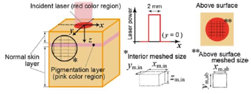
In their study, Dr. Kono and his team considered a simplified skin model consisting of two layers, with a ‘pigmentation layer’ containing many melanosomes, and a ‘normal layer’ containing some melanosomes without any pigmentation (see figure). They assumed the heat absorbed by melanosomes to diffuse to the surrounding tissues via conduction and determined the radiative properties of the pigmented layer from the number density of melanosomes, using it to calculate the energy absorbed per melanosome during irradiation.
The team found that the distribution of the absorbed laser energy and the temperature rise of the skin tissue was indeed influenced by the degree of pigmentation, as anticipated. Specifically, an increasing volume fraction of melanosome (ratio of melanosome volume to the whole skin layer volume) resulted in a dramatic decrease in the distribution of absorbed energy per melanosome in the pigmentation layer but only slightly so in the normal layer. The team attributed this to the increase in melanosome number density for a high volume fraction that, in turn, caused a decrease in the absorbed energy per melanosome.
Further, for high volume fractions, the separation between the melanosomes was smaller, which did not allow for sufficient heat dissipation inside the skin tissue and, consequently, caused a gradual increase in the temperature between the melanosomes. In addition, the team found that skin layers deeper than the pigmentation layer showed a higher temperature compared to the pigmentation layer. Thus, the team concluded that a higher degree of pigmentation (darker skin) was more prone to suffering damage from laser irradiation.
While these findings provide some interesting insights, Dr. Kono is cautious in his optimism. “I believe that our work is necessary to simulate the laser treatment of skin tissue and thereby improve the safety of the procedure. However, as a next step, it is necessary to verify that our model is consistent with the actual laser heating phenomenon inside skin tissue,” he says.
Source: Read Full Article