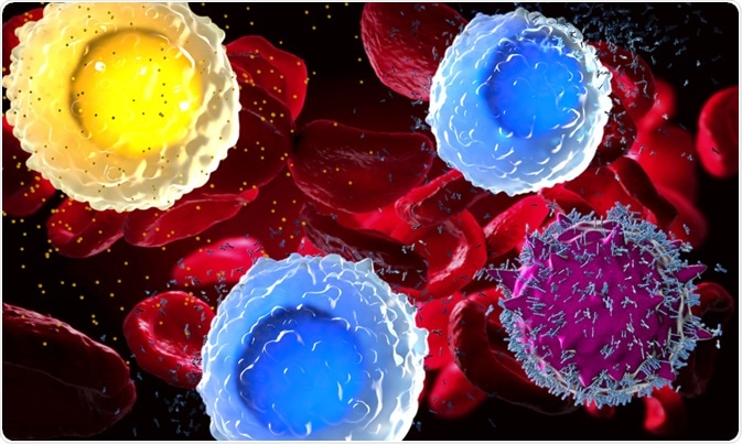
Analyzing peripheral blood mononuclear cells (PBMCs) is an important methodological approach used to evaluate the status of a person's immune system. In short, PBMCs include B cells, T cells, Natural Killer (NK) cells, and dendritic cells.
 Christoph Burgstedt | Shutterstock
How to isolate PBMCs from whole blood
Dilution
Blood can be stored at room temperature before isolating PBMCs, and then remixed by inverting the collection tube 6–8 times. After wiping the tube with 70% ethanol, the contents should be transferred to another tube that can at least hold twice the volume of transferred blood.
Now, the blood can be diluted with an equal volume of phosphate-buffered saline (PBS). In certain cases, Roswell Park Memorial Institute (RPMI) medium is used instead of PBS to increase the viability of cells. The important thing is that the blood and media should be mixed sufficiently.
Separation using density gradient centrifugation
Ficoll PAQUE, a sterile medium, is added to the bottom of the new tube. In case of 6–10 ml of diluted blood, 4 ml of Ficoll PAQUE can be added; whereas 30–35 ml of diluted blood should be diluted with 20 ml of Ficoll PAQUE can be added. The diluted blood is then slowly added to density gradient media and centrifugation is performed for around 20 minutes. The buffy coats should be centrifuged at 1900 revolutions per minute (rpm), while the whole blood should be centrifuged at 1300 rpm.
This should be done without the brakes, as de-acceleration can disrupt the density gradient formation. The centrifuge should not shake during the process, and in such cases, the device should be stopped immediately, reweighed and balanced.
Collecting PBMCs
During the washing of PBMCs, precautions should be taken to ensure that there is no contamination of the platelets. New sterile conical tubes with same volume as density centrifugation should be used to contain the PBMCs and as much of the plasma as possible should be removed.
The monolayer cells from the plasma can be collected using a transfer pipet. These cells can be transferred to a new tube; however, the cells should not be pooled yet. Only plasma should be aspirated during the collection and each tube can then be filled with PBS.
During the first wash, the cells should be spun at 1300 rpm for 8 minutes. After the process, the supernatant can be carefully removed and the pellet can be resuspended in 1 milliliter PBS. In the second wash, the solution should be centrifuged at 1300 rpm for 8 minutes, the supernatant is removed, and the pellet resuspended in 1 mL PBS.
After performing another wash step, the cells from the same donor can be pooled, and to remove platelets, the solution is centrifuged at low speed of 1000 rpm for 10 minutes. This step can be removed if the volume of the blood is low.
PBMC count and viability
After the last step, the supernatant can be removed and the pellet resuspended in 1mL PBS. PBS can be added such that the concentration is 5 X 10⁶ cells/mL. Using cellometer, the cell concentration and viability can be determined.
The last step necessitates staining cells with acridine orange (which stains nuclei) and propidium iodide (in order to stain compromised cells with damaged membranes). For this, 20 µL of cells can be mixed with 20 µL of acridine orange/propidium iodide. After mixing 5 times and loading in to the chamber, the live cell and total cell concentration can be recoded on the machine.
PBMC freezing
The cells can be spun at 1300 rpm for 8 minutes and, subsequently, the supernatant can be removed. The pellet can be resuspended in cold FBS at a concentration of 4 X 10⁷ cell/mL.
The cells can be further detached/removed by further gently pipetting with FBS. Then, same amount of cold FBS is added dropwise while gently mixing the cell suspension. After mixing by gently pipetting, 1mL of this solution can be added to a cryotube and frozen.
Sources
- www.cptp.inserm.fr/…/PBMC-isolation-and-cryopreservation.pdf
- www.ncbi.nlm.nih.gov/pmc/articles/PMC4673925/pdf/ijpr-14-0979.pdf
- www.gelifesciences.com/…/ficoll-paque-plus-density-gradient-media-p-05824
Further Reading
- All Hematology Content
- What is Hematology?
- What is Hematopoiesis?
- Hematology Tests
- Hematology Treatments
Last Updated: Jan 3, 2019

Written by
Dr. Surat P
Dr. Surat graduated with a Ph.D. in Cell Biology and Mechanobiology from the Tata Institute of Fundamental Research (Mumbai, India) in 2016. Prior to her Ph.D., Surat studied for a Bachelor of Science (B.Sc.) degree in Zoology, during which she was the recipient of anIndian Academy of SciencesSummer Fellowship to study the proteins involved in AIDs. She produces feature articles on a wide range of topics, such as medical ethics, data manipulation, pseudoscience and superstition, education, and human evolution. She is passionate about science communication and writes articles covering all areas of the life sciences.
Source: Read Full Article