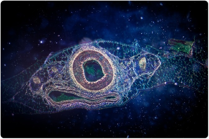
Micropatterning is the creation of specifically patterned and textured surfaces to study the effects of the cellular microenvironment on cell, tissue, or organ behavior.
 Pan Xunbin | Shutterstock
Micropatterning allows for greater control over factors such as spatial constraint and chemical exposure in a three-dimensional platform. This not only allows for the study of cells exposed to these circumstances but also the development of microfluidic devices such as assays.
Micropatterned devices have already been developed as replacements for some in vivo animal models. They are able to replicate the conditions of a particular tissue or organ better than traditional petri dish methods, by forcing the cells to adopt a shaped pattern of adhesion.
How can cells be engineered by micropatterning?
The microenvironment created by micropatterning allows for several physical parameters to be controlled in three dimensions. The volume of particular compartments can be fine-tuned in order to limit cell size, shape, and ability to divide, in addition to the orientation and level of contact with adjacent cells, and the shape of the extracellular environment. The latter factors are important when studying signaling between cells (intracellular). The finely patterned texture of the compartment also plays a role in cell adhesion.
The three-dimensional architecture of micropatterned wells is one of the most useful features when studying cell behavior, as cells forced to grow on a two-dimensional substrate may behave significantly differently than they would in a natural environment. For example, breast cancer cells frequently form unnatural islands of cells across a petri dish surface.
The material from which the micropatterned substrate is made may also play a role in cell behavior. Soft-walled wells force cells to provide themselves with a greater degree of rigidity through the greater expression of cytoskeletal actin within the cell.
Controlling the cytoskeletal growth of cells is possible by using micropatterns in a wide variety of shapes, including spheres, triangles, and elongated rods, among others.
Controlling the cytoskeleton is useful for cell biologists, as extracellular protein complexes on the outer surface of the cell membrane tend to form preferentially at distal sites such as the corners of a triangular cell, known as focal adhesions. This leaves the flat surfaces of the cell membrane less populated with such protrusions, and so makes the study of specific receptors or membrane penetrating agents simpler.
Creating shaped cells also allows cell behaviors such as migration and cell division to be studied. For example, tear-drop shaped micropatterns cause cells to form a denser actin network towards the curved side and tend to migrate in that direction due to polarity effects. It is critical to characterize the effect of cell shape on migration and division is critical when studying cancer cells.
What are the practical applications of micropatterning in biology?
micropatterning allows small assays to be developed that are highly transportable and convenient. These are therefore known as ‘lab on a chip’ systems. Multiple analytes may be tested on a single chip, and detected by a variety of methods including fluorescent staining, in real time. Only very small volumes of sample are needed, and can be drained from the chip at any point for re-use of the assay.
How are micropatterned materials made?
The most common methods of creating micropatterns on materials are variants of lithography, several of which exist. Generally, lithography is done by coating a solid material substrate with a material that can be easily removed by chemical or physical means, in the shape of the desired pattern.
The substrate is then firstly exposed to one chemical or physical process such as acid etching, UV-light, or an electron beam, which is resisted by the coating, followed by a second exposure to a chemical or physical operation to remove the coating. This leaves the desired pattern imprinted on the substrate.
Source
- Methods of Micropatterning and Manipulation of Cells for Biomedical Applications.
- Micropatterning as a tool to decipher cell morphogenesis and functions.
- Micro-patterned porous substrates for cell-based assays.
- Micropatterned surfaces: techniques and applications in cell biology.
Further Reading
- All Micropatterning Content
Last Updated: Feb 27, 2019

Written by
Michael Greenwood
Michael graduated from Manchester Metropolitan University with a B.Sc. in Chemistry in 2014, where he majored in organic, inorganic, physical and analytical chemistry. He is currently completing a Ph.D. on the design and production of gold nanoparticles able to act as multimodal anticancer agents, being both drug delivery platforms and radiation dose enhancers.
Source: Read Full Article